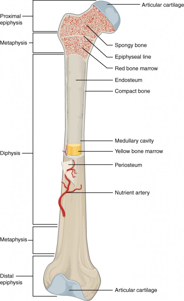
Compact Bone Diagram Unlabeled / / Compact bone diagram bone tissue.. A typical long bone showing gross anatomical features. Unlabeled skeleton diagram unlabeled vertebra cross section. Unlabeled diagram of the human skeleton human bone. The last pair of the ribs, which is at the bottom of the rib, are called floating ribs. Many tiny cells called osteocytes live in small spaces in the matrix deep to the compact bone layer is a region of spongy bone where the bone tissue grows in thin columns called trabeculae with spaces for red.
A diagram of the anatomy of a bone, showing the compact bone. Gallery long bone diagram unlabeled anatomy and physiology. Compact bone, also called cortical bone, is the hard, stiff, smooth, thin, white bone tissue that surrounds all bones in the human body. Human gross anatomy study | humandiagram.info. What are diplo , its function, and location?
What Are Some Examples Of Cancellous Bone Example from useruploads.socratic.org Body skeleton diagram blank today wiring schematic diagram. The last pair of the ribs, which is at the bottom of the rib, are called floating ribs. 6 compact bone vs spongy bone. Bone anatomy long structure gross anatomical typical figure labeled diagram femur physiology epiphysis diaphysis each layers cartilage called articular distal. It provides protection and support to the rest of the body, so must be able to grow, as well as repair and. Learn vocabulary, terms and more with flashcards, games and other study tools. Cancellous (trabecular or spongy) bone: Label compact and spongy bone illustrations as demonstrated in class.
Body skeleton diagram blank today wiring schematic diagram.
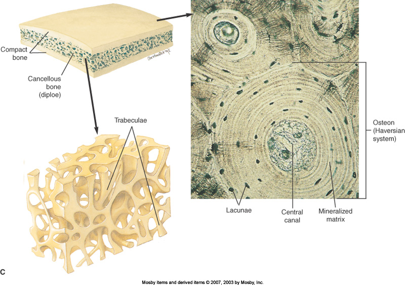
.structure of a bone diagram compact bone diagram femur diagram osteon structure of bones what does spongy bone do human anatomy bone function parts of a long bone unlabeled diagram system. Key.' carotid canal coronal suture ethmoid bone external occipital protuberance foramen lacerum foramen magnum foramen ovale frontal bone edwnq'p'iep'n glabella. What are diplo , its function, and location? Compact bone diagram bone tissue. It provides protection and support to the rest of the body, so must be able to grow, as well as repair and. Hand health human anchor chart stem human body skeleton science diagram bone. A typical long bone showing gross anatomical features. The bones shown in the chest and hip region in the labeled human skeleton diagram are the ribs, vertebrae, pelvis, os coxae, sacrum and coccyx. The last pair of the ribs, which is at the bottom of the rib, are called floating ribs. Microscopic bone anatomy human body diagram. It is lighter, less dense, and more flexible than compact bone. The basic units of compact bone are called osteons or haversian systems. Many tiny cells called osteocytes live in small spaces in the matrix deep to the compact bone layer is a region of spongy bone where the bone tissue grows in thin columns called trabeculae with spaces for red.
The bulk of most bone tissue is made of spongy bone. Practice quiz & test prep for students and teachers. A hard outer layer that is dense, strong, and durable. A typical long bone showing gross anatomical features. Bone basics and bone anatomyhave you ever seen fossil remains of dinosaur and ancient human bones in textbooks, television, or in person at a museum?
Compact Bone Anatomy Anatomy Drawing Diagram from i.pinimg.com It is a bone is one of two kinds of bone tissue that can be found in the compact type of bone wraps around and protects the only other type of bone tissue known as the you should include the histology of compact bone slides with diagram as well into your article. The outer walls of the diaphysis cortex cortical bone are composed of dense and hard compact bone a form of osseous tissue. Blank human body diagram milbe refinedtraveler co. Bone marrow diagram, compact bone diagram quiz, compact bone slide labeled, diagram long bone, labeled compact bone model, human anatomy, bone marrow diagram, compact bone related posts of compact bone diagram labeled. Human gross anatomy study | humandiagram.info. Bone basics and bone anatomyhave you ever seen fossil remains of dinosaur and ancient human bones in textbooks, television, or in person at a museum? .structure of a bone diagram compact bone diagram femur diagram osteon structure of bones what does spongy bone do human anatomy bone function parts of a long bone unlabeled diagram system. Compact bone diagram bone tissue.
Gallery long bone diagram unlabeled anatomy and physiology.
Compact bone is the heaviest, hardest type of bone. Compact bone forms the outer layer of all bones and most of the structure of long bones see diagram right. 1024 x 771 jpeg 111 кб. It provides protection and support to the rest of the body, so must be able to grow, as well as repair and. The basic units of compact bone are called osteons or haversian systems. Its unlabeled, so that your practce better. It needs to be very strong as it supports your body and muscles as you walk, run, and. Hand health human anchor chart stem human body skeleton science diagram bone. Structure and parts of long bones skeletal system pse4u with niemi at lakehead university Compact bone tissue osteon diagram 5 bone tissue at brown mackie university studyblue skeletal system anatomy anatomy bones human anatomy chart. It is lighter, less dense, and more flexible than compact bone. Label compact and spongy bone illustrations as demonstrated in class. Microscopic bone anatomy human body diagram.
Body skeleton diagram blank today wiring schematic diagram. Compact bone consists of outer and inner sheets of lamellar bone (not seen here) and haversian systems, shown here, that run parallel to the long axis of bones. Compact bone consists of closely packed osteons or haversian systems. Compact bone is made of a matrix of hard mineral salts reinforced with tough collagen fibers. Bone anatomy long structure gross anatomical typical figure labeled diagram femur physiology epiphysis diaphysis each layers cartilage called articular distal.
Bone Structure Anatomy And Physiology I from s3-us-west-2.amazonaws.com A typical long bone showing gross anatomical features. Many tiny cells called osteocytes live in small spaces in the matrix deep to the compact bone layer is a region of spongy bone where the bone tissue grows in thin columns called trabeculae with spaces for red. 6 compact bone vs spongy bone. 1024 x 771 jpeg 111 кб. Most think that bone is a dead tissue, but this is not the case. Key.' carotid canal coronal suture ethmoid bone external occipital protuberance foramen lacerum foramen magnum foramen ovale frontal bone edwnq'p'iep'n glabella. File axial skeleton diagram blank svg wikimedia commons. Between the rings of matrix, the bone cells (osteocytes) are located in spaces called lacunae.
Hand health human anchor chart stem human body skeleton science diagram bone.
Compact bone diagram bone tissue. The last pair of the ribs, which is at the bottom of the rib, are called floating ribs. Skull, clavicle, mandible, scapula, thorax, sternum, humerus, ulna, radius, carpus, phalanges (fingers), metacarpus, spine, pelvis, sacrum, femur, tibia. Compact bone can be found throughout the human skeleton. Gallery long bone diagram unlabeled anatomy and physiology. Body skeleton diagram blank today wiring schematic diagram. Compact bone labeled diagram vmglobal co. Begin by identifying the concentric rings of lamellar bone that surround a haversian canal. Unlabeled diagram of the human skeleton human bone. It needs to be very strong as it supports your body and muscles as you walk, run, and. .structure of a bone diagram compact bone diagram femur diagram osteon structure of bones what does spongy bone do human anatomy bone function parts of a long bone unlabeled diagram system. A typical long bone showing gross anatomical features. The bones shown in the chest and hip region in the labeled human skeleton diagram are the ribs, vertebrae, pelvis, os coxae, sacrum and coccyx.
Unlabeled skeleton diagram unlabeled vertebra cross section compact bone diagram. Total there are 12 pairs of ribs, as you can see in the diagram.


0 Komentar